Feeds:
Posts
Comments

Archive for the ‘Fungus growth 2013’ Category

Comatricha nigra is a new slime mold to me, but is one of the more common species in the UK. It grew on a piece of ash branch that has been brought down in the recent high winds, the branch then lay on a path being drenched by heavy rain on approximate alternate days for a while before I brought it home where I kept it in the dry. Then, unexpectedly, white jelly-looking rounded growths emerged. They became pink, then grew black stalks and became darker pink, then brown and finally black, and were shiny all the time. They are very small only 2 to 3 mms high.

Comatricha nigra, just emerged, white and stalkless, 8.8.2013.

Comatricha nigra, black stalks and maturing through pink to brown. 10.11.2013

Comatricha nigra, black and shiny, 11.11.2013

Details of the growth are in my notes.

Read Full Post »

There have been a lot of articles written about fungus foraging, what damage it does etc. I am going to add my ideas with regard to the situation in Abney Park.

Fungal fruiting bodies (FBD) are the equivalent to a flower, and they sit on and are produced by hyphae. The hyphae are the main body of the fungus. Have you ever kicked over damp leaves and seen a white fibrous matter linking the lower leaves? That is hyphae. Occasionally it is thick enough to be visible as in the ‘bootlaces’ of honey fungus, renowned for the black mesh that is seen under the bark when a tree has died. Removing the flower from a plant can stimulate more flower production without really damaging the plant. Removing the FBDs from the hyphae might also stimulate the production of more FBD, it might exhaust the whole fungus but it is probably going to be less damaging than the compression of the soil around the FBDs which can damage the fine threads of the hyphae.

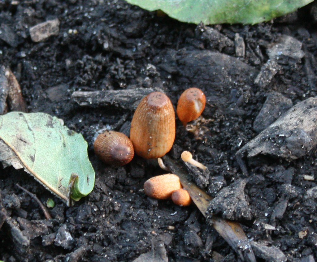
Bootlaces of Armillaria, 1.2.2013

The impact on the soil in Abney is minimal as most walking is directed along paths and the off path traffic is limited by the graves.

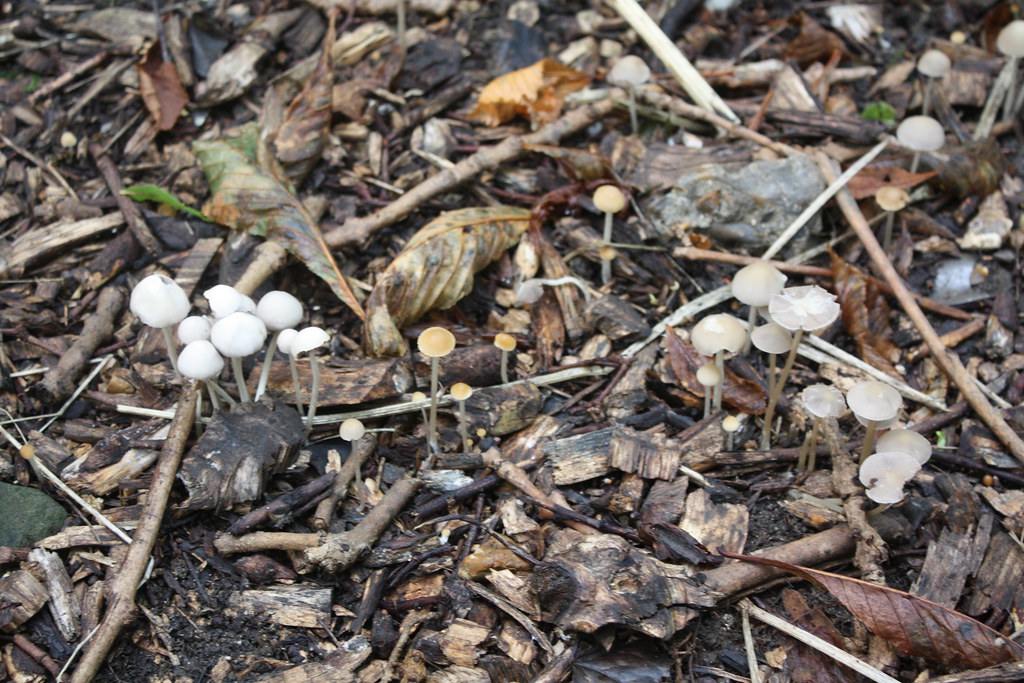
What could be more annoying is the effect on the nature reserve of removing or prematurely knocking over clusters of caps. Agaricus and Chlorophyllum are the prime species involved here. Each year I find clusters of caps picked and discarded. They have not been kicked over but are free of the ground and on their sides around where they have been picked. Agaricus is a group of species that includes edible types, but in Abney they are probably not. They conform to the “if I can peel it and it has a white cap it is safe to eat” that has been quoted at me occasionally, but it doesn’t work. Most Agaricus caps in Abney are yellow stainers which the books will tell you cause projectile vomiting etc. The best way to tell is by picking a fresh cap and bruising the base… and watching it go bright yellow. If a cap from one cluster goes yellow the chances are that all the caps in the immediate area will do the same. So why pick a whole group? If you see anyone doing this would you ask them for me? At least they still produced spores when they are maturing in the place where they grew.

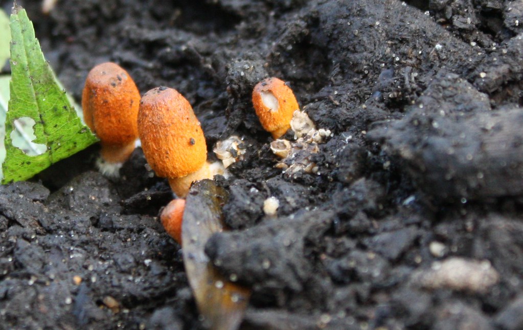
What is more interesting to me is the invertebrates that are associated with the caps. I have spent a lot of time recently looking at the little creatures associated with them. Yesterday I looked at an exuberant flowering of Glistening Ink Cap, one of the more common ink caps in Abney. There were 2 flying insects living around the caps. There was a Fungus Gnat, and I am going to quote Wickipedia here….

Fungus gnats are small, dark, short-lived flies, of the families Sciaridae, Diadocidiidae, Ditomyiidae, Keroplatidae, Bolitophilidae, and Mycetophilidae (order Diptera); they are sometimes placed in the superfamily Mycetophiloidea. The larvae of fungus gnats feed on plant roots and fungi, which aids in the decomposition of organic matter. The adults are 2–5 mm long and are important pollinators that can help spread mushroom spores as well as plant pollen.

Coprinellus micaceus, Glistening Ink Cap 27.10.2013 with fungus gnat

And then there was a more substantial fly that I have yet to get even close to identifying.

Coprinellus micaceus, Glistening Ink Cap 27.10.2013 with fly

A small spider was living under the rim of one of the caps and making a very fine living from darting out and grabbing the odd gnat. I’ve seen a robin and a wren feeding on these flies.

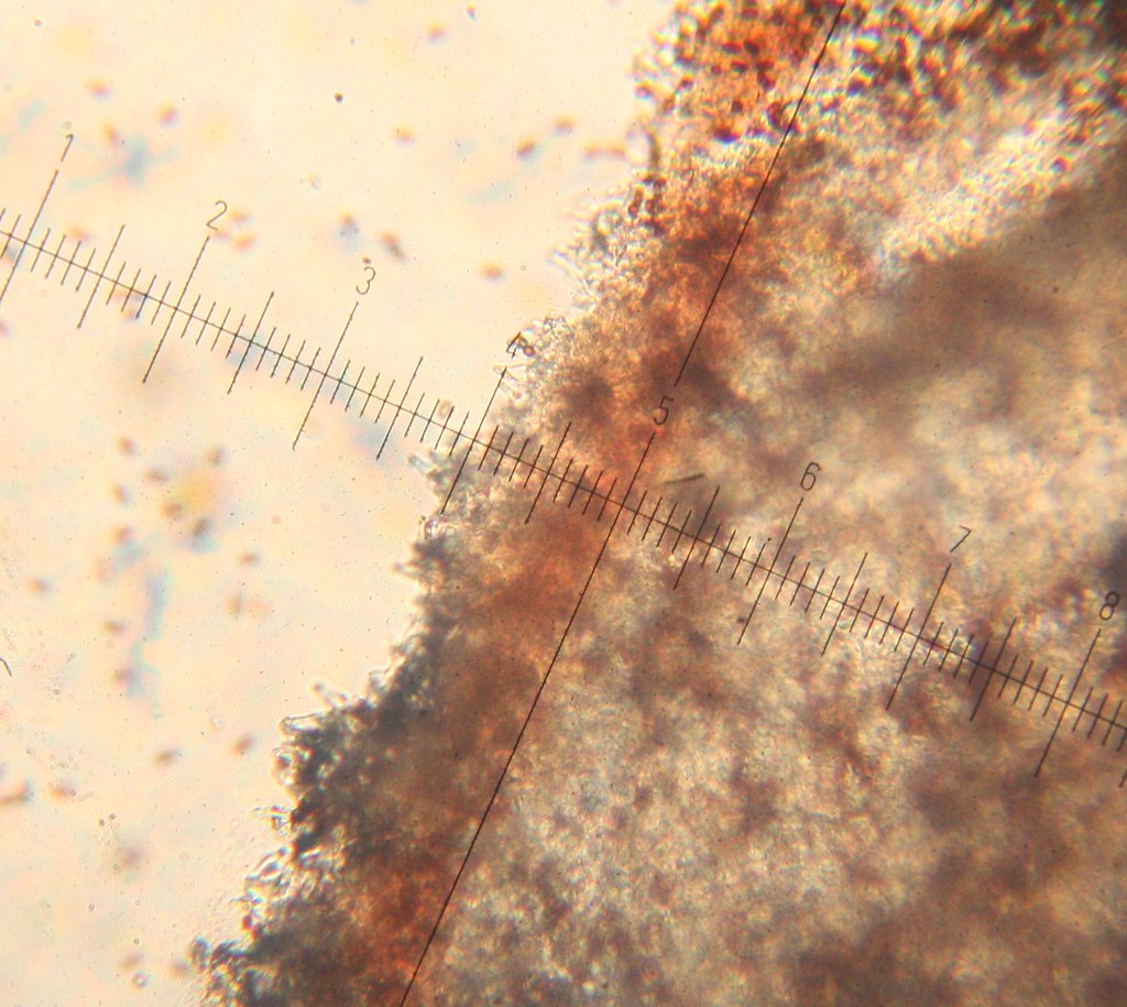
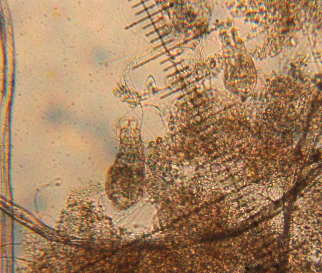
When I look at slides taken from FBD to try to identify them I continually coma across microscopic animals. Apart from the maggots there are even smaller foraging creatures I have no idea about. There are a couple of examples here but they are the tip of the iceberg…

Invertebrates feeding in cap 20.10.2013 (1)

Invertebrate feeding in cap (2)


If the fungus caps are removed or prematurely shattered (stamping) or dried (picking and leaving on the ground) it is taking out a section of the network of interrelations that are important in a nature reserve.

Read Full Post »

There are only 45 records of Coprinopsis erythrocephala in the British Fungi Checklist, so it doesn’t grow very commonly. It is growing happily in Abney at the moment, on soil just in a vulnerable place where it can be trodden on so easily along the north boundary path, so I’ve got in quick with collecting and making notes.

Caps when first seen, 8.10.2013

Caps 10.10.2013

It is not a mature growth that I’ve described, but it does have all the right features already in place. The most distinctive feature is the colour of the young caps, they are orange/red. This fades as they mature to become blackish. Once that orange shows up there is little else that it can be. I’ve put my notes here.

Read Full Post »

I find this interesting (and thanks to Russell for pointing it out to me). Pluteus aurantiorugosus is a rare species that grows in Abney every year on a large number of sites, but always on dead wood.


Here it is growing on the side of a live Common Ash tree. This indicates that the wood directly round the cap is dead. It has been killed by the bracket – Aurantioporus fissilis. I followed the progress of another outcrop of this bracket in another blog, and it does look as if the wood has suffered around the edge. There is no way of knowing that the wood around the hole is not being inhabited by the bracket that created it. This finding suggests that the Aurantioporus lives just in the heart wood, and degrades only the wood that is in its way to the outside where it can shed its spores. This leaves the dead wood around the hole available to be inhabited by another fungus species, in this case the Pluteus aurantiorugosus.

Read Full Post »

Along one particular pathway are a multitude of small caps, and they all look a little bit different and they all end up in the key as being the same, Psathyrella pseudogracilis. The colour is variable, they begin red/brown, and become yellowish (sometimes) or a watery grey, and that’s just when they are fully hydrated. When they dry out they are opaque yellowish cream or pinkish cream, depending on if they started yellowish or red/brown. If they dried from grey they turn chalky white. They can grow singly or in tight clusters so they fuse at the base.

Psathyrella pseudogracilis on buried wood, dry

Psathyrella pseudogracilis, traditional hydrated grey with radial lines,


Young red/brown Psathyrella pseudogracilis, one drying in the centre so becoming paler.

Clusters of Psathyrella pseudogracilis, dry on the left, less dry on the right.

So why are they all the same species? They all grow on wet wood or wood chips. They all have gills that start out palid with a light edge, then develop a white edge underlined with red. They all have the same varieties of cheilocystidia (the large white edge cells along the gills) and pleurocystidia (slightly larger but essentially similar cells on the gill surface. They smell the same, a bit musty. The spores are all black and the same oval shape with a little off-center tail and central germ pore, and all are in the right size range for the species, 11 to 16 microns long and 6 to 7.5 microns wide. This is a group where a microscope is really essential.

Squash slide of gill edge of Psathyrella pseudogracilis, with the cheilocystidia slightly loosened to show a couple of whole cells.

The white edge with red below of the gills of Psathyrella pseudogracilis under a microscope.

Although they are fading a bit now it is still worth a look at path edges for them, as when they do grow they make up for their small size in the hundreds that can grow at one time.

Link to my notes is here.

Read Full Post »

The most fantastic growth of caps I have yet seen developed on a collapsed poplar tree.

The tree was degraded by Ganoderma australe from the ground to almost branch level, and in the hollow trunk at branch level Rigidoporus ulmarius. Ganoderma australe causes white rot, ie biodelignification of wood. Rigidoporus ulmarius causes brown cubical rot of trees, ie lignocellulose breakdown of wood. These are the two strengthening components of wood and without them the wood has degraded into a soft, spongy texture. Not surprisingly it collapsed.  The fallen trunk recently absorbed water and provided the perfect food for the Volvariella bombycina to grow.

The name Volvariella refers to a volva, a sac that the cap developes inside, which then breaks at the top as the cap grows out of it. The stem is without a ring. The gills start out pale, then become pink (rose), and eventually brown. The cap is covered in hairs, so ‘silky’.

It is a rare cap, with 314 records held at Kew for the UK.

Link to notes of Volvariella bombycina.

Link to photos of fungi and slimemolds 2013 in Abney Park Cemetery.

Read Full Post »

There it was, on the old rotten trunk, and one cluster of growth was a bit more exposed, just a bit wetter than the others. It was reverting to plasmodium feeding stage. This is the first time I have seen the bright red plasmodium. It looks a bit of a mess here, but this is the amoeba like body that moves through the wood in search of food. It moves to the surface when the conditions are right, and there changes form to become the vivid orangey-pink round aethalia, which produce the spores. All my notes are here.

Read Full Post »

Just occasionally I feel a bit sad to find a new species of fungus… and this is one of those. The Cherry Plum with its distinctive purple foliage near the Church Street entrance is now infected with Taphrina pruni. It has got at the vast majority of the developing fruits and they are now sterile, enlarged and powdery. The fruits would usually feed many birds and insects etc so this is a loss for further into the year. It is worth a look for the oddness of these strange heavy galled fruits hanging on the ends of the lower branches. Notes are here.

The pocket plums were not discovered by me, but on a walk about foraging given by Nick from Hodmedods, a company selling British peas and beans that have grown in Britain since the ice age. It was a most enjoyable walk.

Read Full Post »

Kretzschmaria deusta, Brittle Cinder, grows on rotten stumps and logs. and is also a plant pathogen. It kills trees and lives on in the wood. It breaks down lignin and cellulose, the structural strengthening materials in broad-leaved tree wood.

Abney at the moment has a series on log piles augmented by imported wood brought in by tree surgeons. Kretzschmaria deusta fruiting bodies are covering these log piles. This particular log is interesting. The black lines running through the sawn section are zone lines, tree defences. The area around the fungal infection is cut off from the rest of the tree by a waterproof layer that includes melanin, so appears black. If too much of the trunk is blocked the tree can’t transfer water up the tree or the sugars etc from the photosynthesising leaves back down to the roots, and the tree dies. In cross-section these waterproof layers can be seen as black lines.

In this photo the zone lines are around a section of the cut log. Inside the area the wood is wetter and more degraded than outside. The Kretzschmaria deusta fruiting bodies, the flat grey growths with a white edge, have developed on the surface. This is probably the fungus that caused the tree to end up in Abney as logs. It is not often that the logs give away the secret of which fungus has invaded them.

Read Full Post »

Wood plays a huge part in the fungi in Abney, and the inkcaps are specialists in dead wood, so it is not surprising that inkcaps are among the first caps to grow fresh this year.

These were the frist new caps I saw. Glistening ink cap, Coprinellus micaceus), the most widespread of the inkcaps, orangey brown with light crystalline specks on the top.

Then there is the Coprinellus truncorum, only 114 British Records held at Kew and only on one rotting fallen tree in Abney. It looks like C. micaceus with the micaceus specks washed off the top (which can happen easily in light rain).

It takes a microscope to tell them apart.

The smallest of the inkcaps was just beginning yesterday. It grows on wood and buried wood so looks like it is growing in the ground. Coprinellus disseminatus, now officially ‘Fairy Inkcap’, but I prefer the old common name of ‘Trooping Crumble Cap’ of Fairies Bonnets’.

On woodchip piles there are 2 inkcaps. One, Coprinopsis lagopus, is traditional inkcap; transparent grey lined cap that self digests to spread its spores, eventually leaving only the white stalks. It appears as scale covered bullet-shaped, poking through the wood chips.

The last of the inkcaps is not the usual presentation. Coprinopsis marcescibilis I always have trouble with this one. It doesn’t self-digest. It is hygrophanous, ie it changes colour depending on how wet it is, in dry weather it is opaque cream, in wet weather is grey/brown. It used to be Psathyrella marcescibilis, so it depends on how old a field guide is as to where it appears, and it does look more like a Psathyrella, with light edge to the gills.

The other 2 species I found yesterday are not inkcaps. Pluteus salicinus is a grey cap with radial fibrils on the cap surface and a slight shine as if it has been polished. There is no ring on the stem and the gills begin white and change to pale pink, (carrying a mirror to look under caps works well here).

The other cap is an untidy growth of Polyporus durus, Bay Polypore. The leathery looking surface has a white stem here as it is so new, but it does develop a black base. The underside is also white and with pores instead of gills.

The image on the right of the underside is taken through a mirror.

Link to photos of Fungi in Abney 2013

Read Full Post »